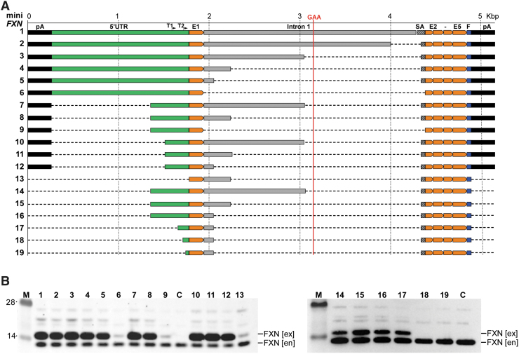
Figure 2.
Transient expression of miniFXN constructs in HEK293T cells. (A) Schematic representation of all 19 constructs generated and tested in this work. All constructs are depicted to scale (shown above top line). Location of GAA repeats is indicated as a vertical red line. Positions of T1 and T2 TSSs are indicated. The 5′UTR sequence upstream of the ATG start codon is shown in green, intron 1 (including splice acceptor sequence, SA) in gray. All FXN exons (E1, E2–E5) are depicted in orange. Flag tag (F) is indicated in blue and polyadenylation signals (pA) in black. (B) All 19 minFXN genes were transfected to HEK293T cells in equimolar amounts and cell lysates were prepared 48 h after transfection. Western blot with FXN-specific antibodies detects both endogenous (en) and exogenous (ex) frataxin, migrating slower due to the presence of C-terminal Flag tag. Numbers above the gel lanes correspond to the miniFXN constructs depicted in (A). C represents untransfected HEK293T cells used as a control. M indicates molecular-weight protein marker (SeeBlue™ Plus2 Pre-stained Protein Standard). Representative images of three independent experiments are shown. miniFXN, frataxin minigene; 5′UTR, 5′ untranslated region.

