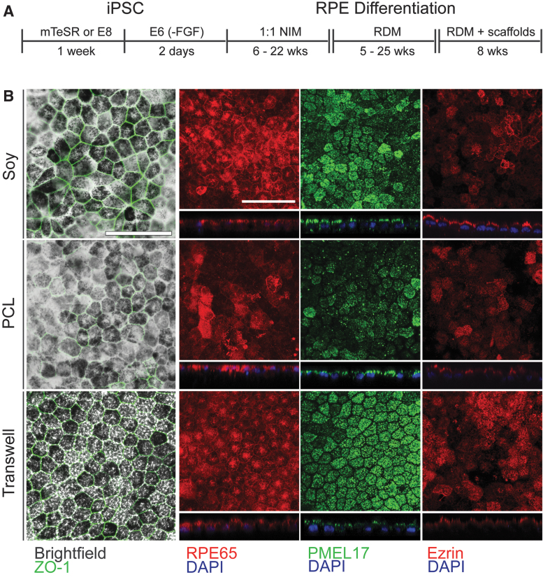
FIG. 4.
Differentiation of RPE on scaffolds. (A) Medium types used over time for the differentiation of iPSCs into RPE. (B) Fluorescence and brightfield microscopy of fully differentiated RPE cells on the three material types. Phalloidin staining and immunostaining using antibodies against PMEL17, RPE65, and ZO-1 are shown as indicated. For all figures, scale bar = 50 μm. iPSC, induced pluripotent stem cell; RPE, retinal pigment epithelium.

