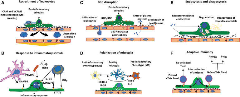
FIG. 3.
Brain pericytes as mediators of neuroinflammation. (A) Recruitment of leukocytes. Pericytes secrete chemokines to actively recruit leukocytes to areas of inflammation. Leukocytes cover gaps between pericytes and utilize ICAM-1 and VCAM-1 guidance to enter the brain parenchyma. (B) Response to inflammatory stimuli. Receptors for PAMPs, DAMPs, and cytokines facilitate pericyte response to pro-inflammatory stimuli. (C) BBB disruption. A variety of factors such as ROS, RNS, and VEGF can promote the breakdown of endothelial tight junctions and basement membrane, leading to BBB disruption. (D) Polarization of microglia. Secretion of pro- or anti-inflammatory mediators by pericytes can alter phenotype for nearby microglia (M1 or M2, respectively). (E) Endocytosis and phagocytosis. Receptors on the membrane detect insoluble materials and dead cells to facilitate their clearance. (F) Adaptive immunity. Internalized antigens from pericytes to primed or naive CD4+ T cells promote T-reg, T cell anergy, or T cell activation. Adapted from Rustenhoven et al.4 PAMP, pathogen-associated molecular pattern; DAMP, damage-associated molecular pattern; RNS, reactive nitrogen species; ICAM-1, intercellular adhesion molecule-1; VCAM-1, vascular cell adhesion molecule-1. Color images are available online.

