Figure 2.

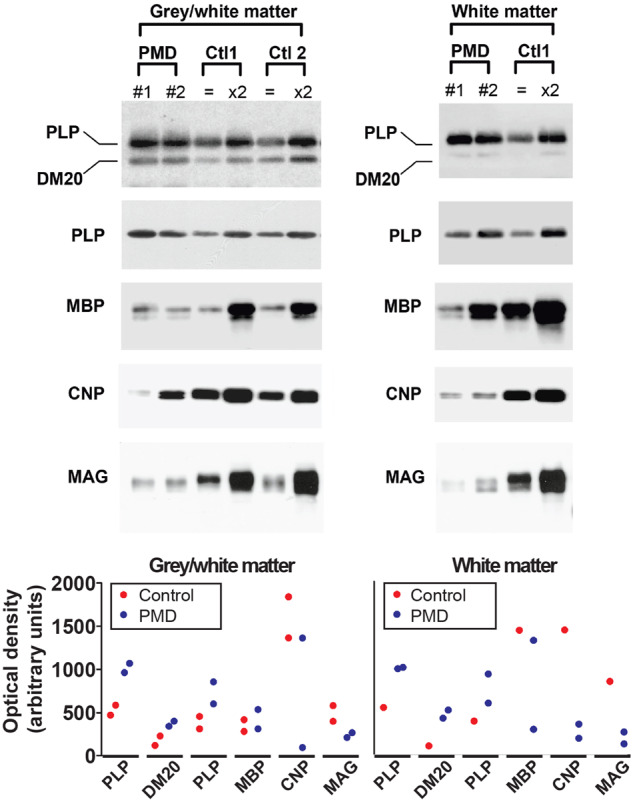
Western blot of myelin extracts from PMD and control human brain show altered levels of myelin proteins. Ctl1 is stored tissue from 70-year-old brain and Ctl2 is fresh biopsy material from 2-year-old brain white matter. Myelin was extracted from tissue that was composed of a mixture of grey and white matter or tissue that was composed almost entirely of white matter. For PLP and MBP analysis, 0.25 μg of the PMD myelin was loaded; in the control patients 0.25 μg (equal symbol) and 0.5 μg (×2) of the samples were loaded onto the gel. For CNP and MAG analysis, 1.5 μg of the PMD myelin and 1.5 μg (equal symbol) and 3 μg (×2) of the control samples were loaded onto the gel. Graphs provide semiquantitative data reflecting band intensities. The y-axis values are the same in the two graphs. Blots were cropped for display purposes. Full-length blots are available in the Supplementary material.
