Figure 4.

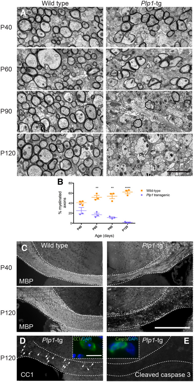
Progressive demyelination in Plp1-tg mouse brain. (A) Electron micrographs of the corpus callosum in wild-type and Plp1-tg mice between P40 and P120. In Plp1-tg mice, at P40 and to a lesser extent at P60, many axons are surrounded by compact myelin, which appears ultra-structurally normal. The proportion of myelinated axons is markedly diminished by P90 and virtually all axons are devoid of myelin sheaths by P120. Astrocyte processes (as) are particularly evident in Plp1-tg corpus callosum at P90 and P120. (B) Graph demonstrating that the proportion of myelinated axons in Plp1-tg mice is similar to wild-type at P40, but diminishes over time. Bars represent mean ± standard error of the mean (SEM). (C) Immunohistochemistry using antibody to MBP at P120 demonstrates the extent of myelin loss across the corpus callosum in Plp1-tg mice. (D) Antibody CC1-positive mature oligodendrocytes are still present in Plp1-tg corpus callosum at P120, suggesting myelin loss is not, or only partially due to the death of oligodendrocytes. (E) Accordingly, most sections of the corpus callosum contained no cleaved caspase 3-positive cells. The positive cell shown in the inset is one of the few cells observed. Scale bars = 2 μm in A; 400 μm in C; 10 μm in inset D.
