Figure 5.

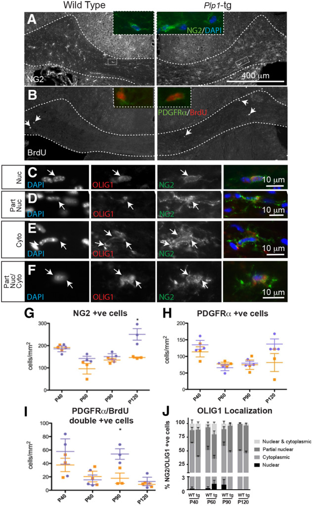
Plp-tg mouse OPCs in adult brain mount a limited response to demyelination. (A and G) NG2 or (B inset, and H) PDGFRα-positive OPCs were present in both wild-type and Plp1-tg mouse corpus callosum at all ages examined, with NG2-positive cells being at slightly but significantly higher density in the P120 Plp1-tg animal compared to the age-matched control (G and H). Wild-type is shown in orange, Plp1-tg in blue. Bars represent mean ± SEM. (B and I) In both Plp1-tg and control animals, a small number of BrdU-positive cells were present in the corpus callosum at 1-h post-BrdU administration. (I) At P90, the number of PDGFRα-positive cells double labelled with anti-BrdU was significantly higher in the Plp1-tg animals compared to control (wild-type in orange, Plp1-tg in blue). Bars represent mean ± SEM. (C–F and J) The basic helix-loop-helix transcription factor OLIG1 translocates to the nucleus during OPC differentiation and the subcellular localization of the transcription factor can be used to assess the status of the OPCs. In Plp1-tg corpus callosum, a small proportion of NG2-positive cells had a nuclear localization of OLIG1 (C and J). Most NG2-positive cells had a partially nuclear (D and J), cytoplasmic (E and J), or partially nuclear and partially cytoplasmic (F and J) localization. Images were adjusted manually for brightness and contrast in Adobe Photoshop for ease of visualization.
