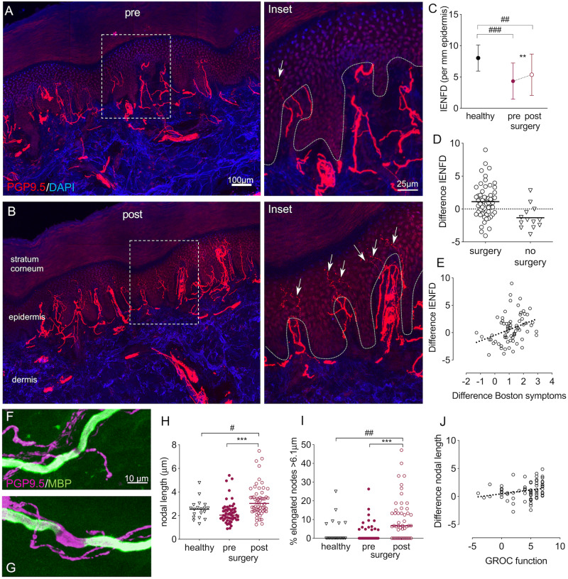
Figure 2.
Histological evidence nerve regeneration in target tissue. Immunohistochemically stained sections of index finger skin of a patient with CTS before (A) and 6 months after surgery (B). Cell nuclei are apparent in blue (DAPI) and axons in red (PGP9.5). After surgery, an increased number of small fibres (arrows on ×4 magnified regions of interest on the right) penetrate the dermal-epidermal border (dotted line). (C) Quantification revealed a reduced IENFD in patients with CTS compared to healthy controls, which increased after surgery but failed to reach normal levels. (D) The difference in IENFD post- compared to pre-surgery demonstrated a high interindividual variability, with a continuing small fibre degeneration in patients who were not operated on. (E) A more pronounced intraepidermal nerve fibre regeneration (difference IENFD) correlates with improved symptoms (r = 0.389, P = 0.001, difference Boston symptom scale data multiplied with −1). (F–J) Changes in nodal architecture. Example of normal (F) and elongated node (G) in skin sections stained immunohistochemically with PGP9.5 and myelin basic protein (MBP). (H) Median nodal length and (I) the median percentage of elongated nodes >6.1 μm increase following surgery and remain higher than healthy controls. (J) The correlation between difference in mean nodal length and the global rating of change scale (GROC) for hand function marginally fails to reach significance after Benjamini Hochberg correction (r = 0.316, P = 0.008). Significant differences for paired/independent t-tests after Benjamini Hochberg correction are indicated between groups with #P < 0.05, ##P < 0.01, ###P < 0.0001 and within groups with **P < 0.01, ***P < 0.0001. Group differences based on n = 59 patients with CTS and n = 20 healthy control subjects.

