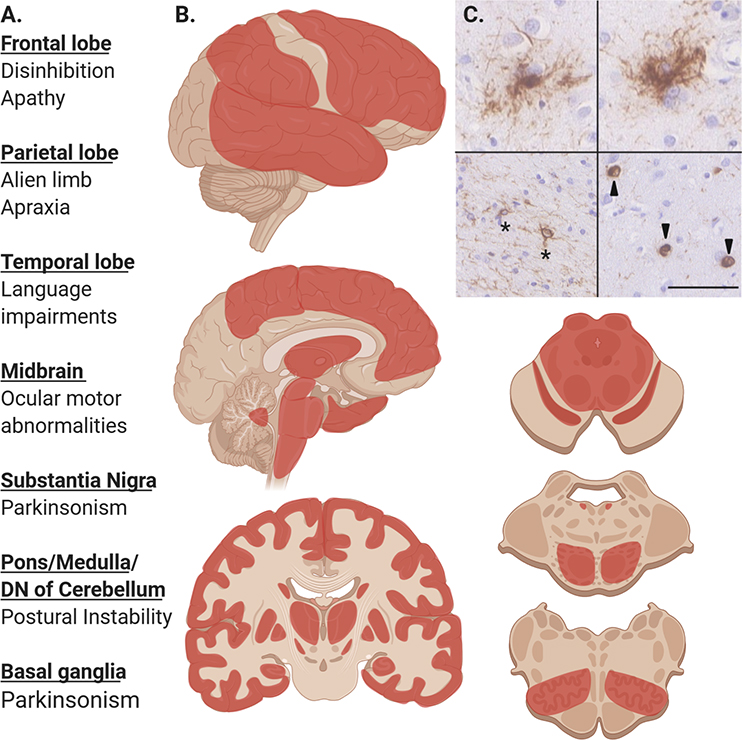
Figure 2: Pathologic Distribution and Clinical Correlations in PSP.
A. Common clinical features of PSP associated with pathology in these affected regions.
B. Regions in red commonly affected by PSP tau pathology and gliosis including, frontal, temporal, parietal lobes, globus pallidus, putamen, caudate, subthalamic nucleus, hippocampus, midbrain tectum and tegmentum, substantia nigra, pontine base and locus ceruleus, inferior olivary nucleus. Darker areas or red are more commonly affected. Certain phenotypes are more likely to exhibit pathology in specific areas (i.e. PSP-SL and temporal lobe pathology and PSP-CBS with parietal lobe pathology)
C. Characteristic microscopic lesions seen in PSP after immunohistochemical staining for phospho-tau with AT8 antibody. Upper row shows two tufted astrocytes, bottom left showing coiled bodies (asterisk), bottom right showing globose neurofibrillary tangles. Scale bar is 50 μm.

