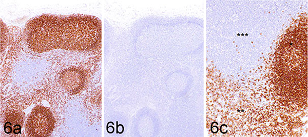
Figure 6.
Normal retropharyngeal lymph node, dog. Immunohistochemistry using the 4E9 mAb. (a) Strong labeling is detected within lymphoid follicles (consistent with B cells) and cells populating medullary cords (consistent with maturing plasma cells). Immunohistochemistry (IHC) for 4E9 mAb without CD19 extracellular domain blocking. (b) No labeling is detected in sections where 4E9 mAb was preincubated with the CD19 immunizing peptide (blocking peptide) prior to its use in IHC. (c) Higher magnification image of panel a. Lymphoid follicle (*), medullary cords (**), and paracortical zone (***).

