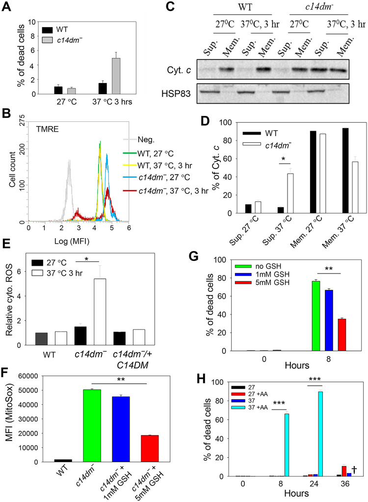
Fig 7. ROS accumulation contributes to heat sensitivity in c14dm‾ mutants.
(A-E) Log phase promastigotes were incubated in PBS at 27 °C or 37 °C for 3 hours. In A, percentages of cell death were determined by flow cytometry after PI staining. In B, ΔΨm was determined after TMRE labeling. Unlabeled WT parasites were included as a negative control (Neg). In C-D, parasites were lysed and cytosolic fractions (Sup.) were separated from mitochondrial fractions (Mem.) as described in Materials and Methods. Distribution of cytochrome c and HSP83 (a cytosolic protein marker) were determined by Western blots (C) and quantified (D). In E, parasites were labelled with the cytosolic ROS indicator DHE. Fluorescence intensities were measured by flow cytometry and relative cytosolic ROS levels were determined. (F) Log phase promastigotes were incubated at 37 °C in the absence or presence of reduced GSH. After 8 hours, mitochondrial ROS levels were measured as described. (G) C14dm‾ parasites were incubated at 37 °C in the absence or presence of reduced GSH and percentages of cell death were determined at 0 and 8 hours. (H) WT parasites were incubated at 27 °C or 37 °C in the absence or presence of AA (10 μM) and percentages of dead cells were determined by flow cytometry at the indicated time points. †: Very few WT+AA cells were detectable after 36 hours at 37 °C. Error bars represent standard deviations from three independent experiments.

