Figure 6.
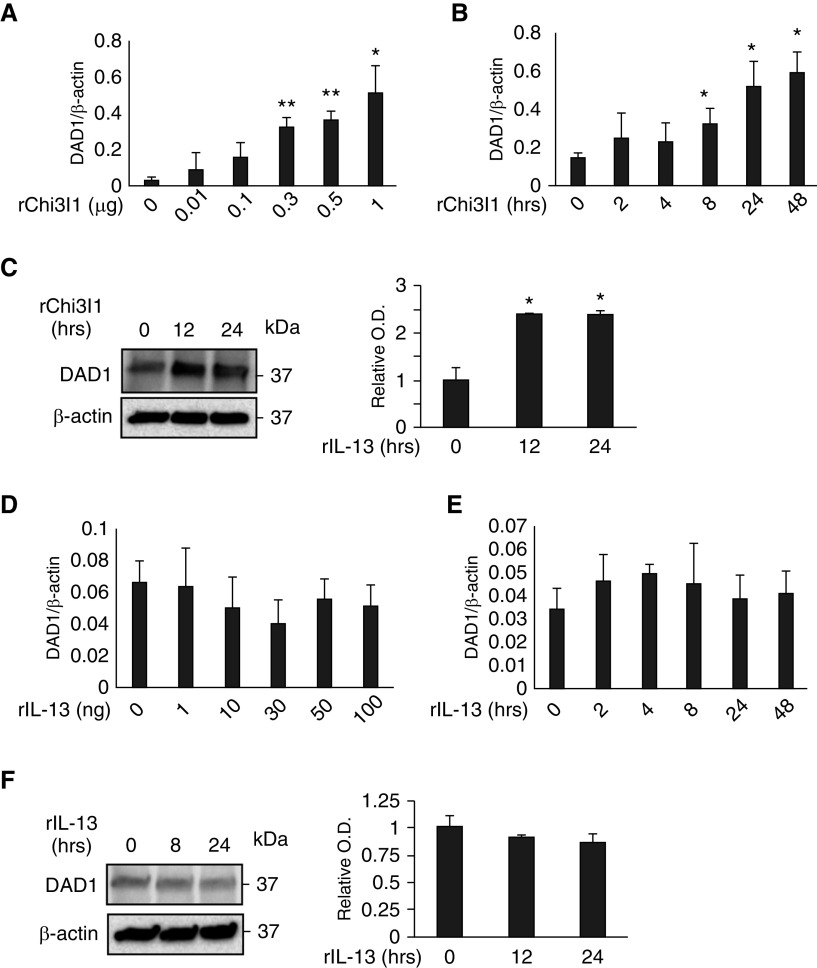
Effects of Chi3l1 and IL-13 on DAD1 (defender against apoptotic cell death 1). MLE12 lung epithelial cells were stimulated with rChi3l1 and rIL-13, then the expression of DAD1 was detected by qRT-PCR and IB assays. (A and B) The mRNA expression of DAD1 gene detected by qRT-PCR in the cells stimulated with different doses (A) and time points (B) of rChi3l1. (C) Representative IBs detecting DAD1 protein expression in the MLE12 cells at different time points of rChi3l1 stimulation. (D and E) The mRNA expression of DAD1 gene detected by qRT-PCR in the cells stimulated with different doses (A) and time points (B) of rIL-13. (F) A representative IB detecting DAD1 protein expression in MLE12 cells with different time points of rIL-13 stimulation. The values in A, B, D, and E represent mean ± SEM in triplicated samples in a minimum of two separate experiments. (C and F) Representative of a minimum of three evaluations. The bar graphs to the right of the IBs in C and F show the relative quantities of DAD1 from three experiments measured by densitometric image analysis. *P < 0.05 and **P < 0.01, compared with vehicle controls.

