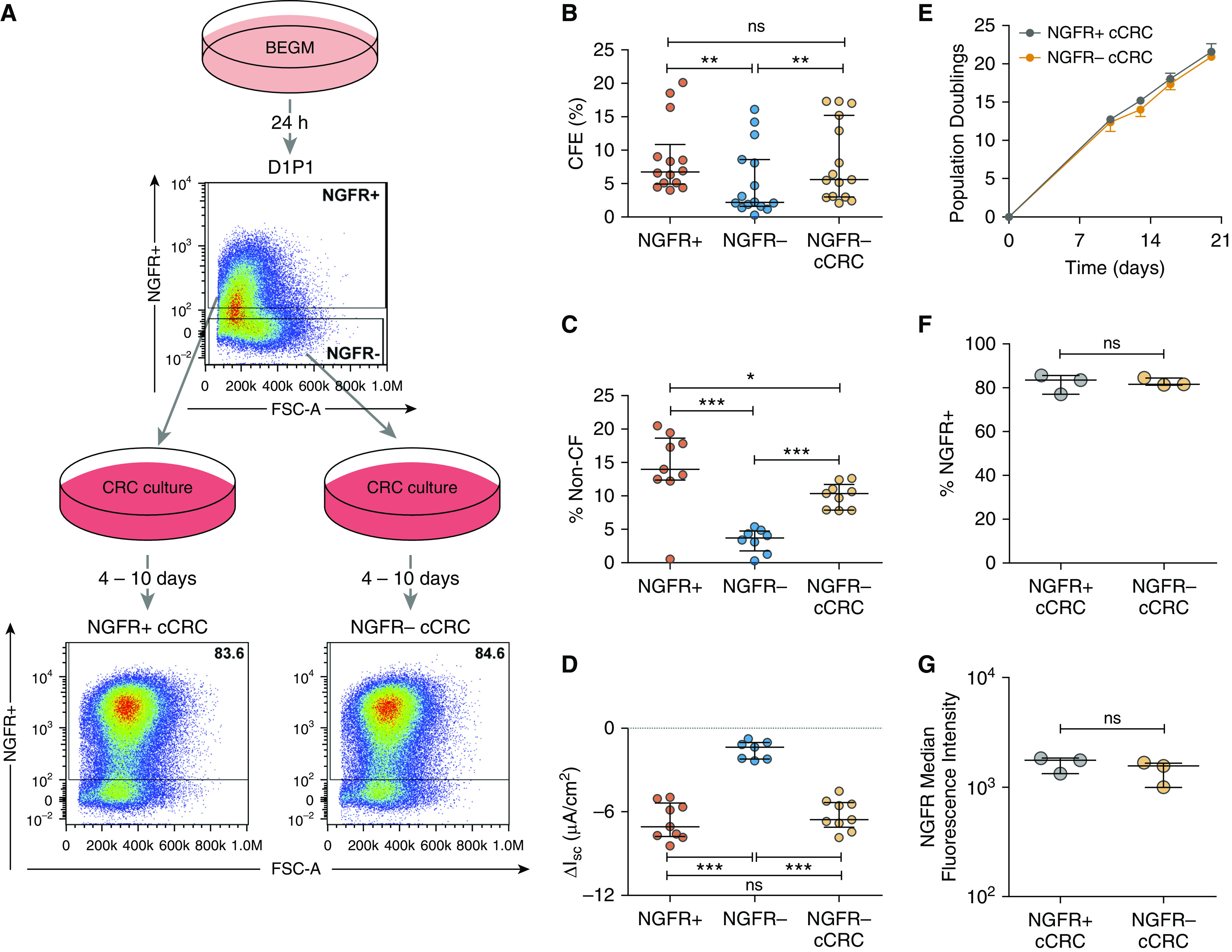
Figure 2.

CRC culture rescues growth capacity of NGFR− human bronchial epithelial cells (HBECs). (A) Schematic outline. Primary HBECs were cultured in bronchial epithelial growth media (BEGM) for 1 day (D1P1), dissociated, stained for NGFR, and sorted based on NGFR expression. Colony-forming efficiency (CFE) and competitive repopulation of NGFR+ and NGFR− cells were analyzed. D1P1 NGFR+ and NGFR− subpopulations were converted to the CRC method (cCRCs), reanalyzed by flow cytometry, and colony forming and competitive repopulation were analyzed. FSC-A = forward scatter area. (B) CFE of NGFR+, NGFR−, and NGFR− cCRCs. Biological n = 5; two to three technical replicates for each donor. Competitive repopulation results: (C) percent non–cystic fibrosis (CF) cells between Days 24–32 (also see Table E3) and (D) short-circuit current response to CFTRinh-172 (CFTR inhibitor-172) in Ussing chambers (also see Table E4). Biological n = 4; one to three replicates per donor. *P < 0.05, **P < 0.01, and ***P < 0.001. (E) Growth curves for NGFR+ and NGFR− cCRCs. Analysis of NGFR expression in cCRCs: (F) percentage of NGFR+ cells and (G) median fluorescence intensity of NGFR in NGFR+ and NGFR− cCRCs. Biological n = 3. Data are presented as median and interquartile range. ΔIsc = change in short-circuit current; ns = not significant.
