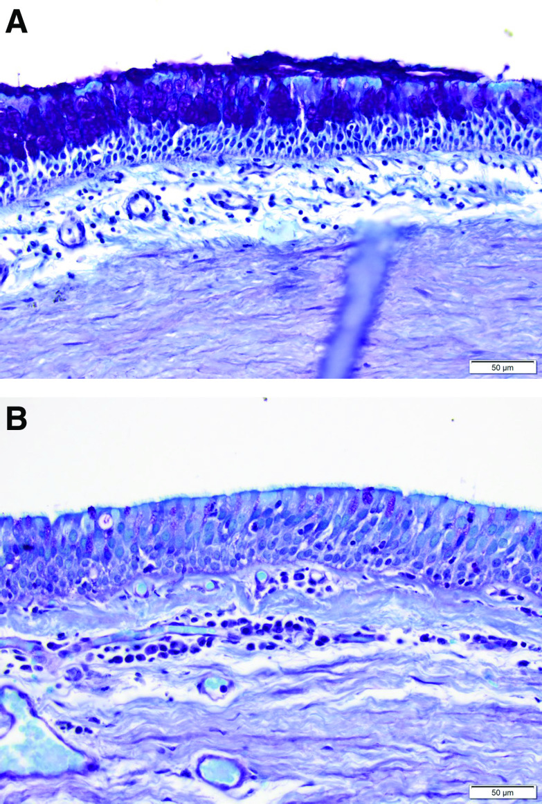
Figure 3.
Histological findings from the right bronchus intermedius of a study patient. The goblet cells, with magenta-colored cytoplasmic mucin highlighted by periodic acid–Schiff staining, are seen in the superficial bronchial epithelium. (A) On Day 0 immediately before therapy, significant goblet cell hyperplasia can be seen (score of 2). (B) Right bronchus intermedius 120 days after the initial treatment, demonstrating complete regeneration of the pseudostratified columnar epithelium with a reduction of goblet cell numbers (semiquantitative assessment score of 1).

