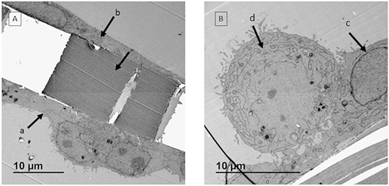
Fig. 4.
TEM images of co-culture model. A: Cell layers with different morphologies were observed on apical side (a) and basal side (b) of the transwell membrane. The space between a & b is the polyester membrane. B: On apical side, two cell layers with different morphologies were detected. The membrane was covered by epithelial cell layer (c) and macrophages were sitting on top of the membrane at an expected ratio (d).

