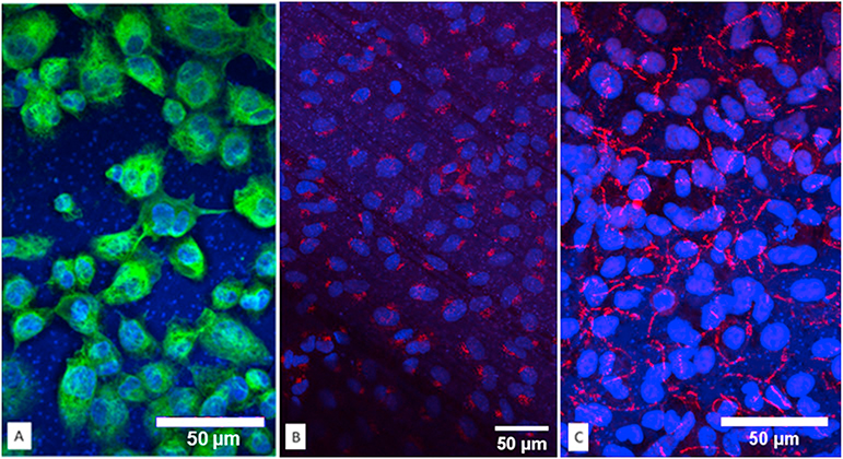
Fig. 5.
Immunofluorescent staining for key protein markers of the co-culture model were detected individually and combined. A: Macrophage CD68 protein signal was observed wrapping around nuclei in green. B: Specific endothelial protein VWF protein was detected only on the basal side of the polyester membranes. C: Tight junction protein ZO-1 lighted in far red as honeycomb shape in A549 cells. (For interpretation of the references to color in this figure legend, the reader is referred to the web version of this article.)

