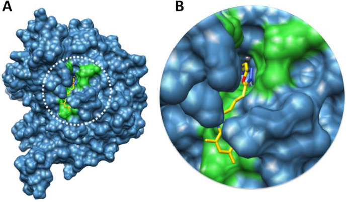
Figure 10.
Docking complex 7e against urease. (A) The urease structure is highlighted in surface format having blue color while the binding pocket is justified in green color in surface format. (B) The closer view of binding pocket which shows the ligand (yellow color) structure and its conformation inside the binding pocket

