. 2020 Aug 11;9:e57081. doi: 10.7554/eLife.57081

Figure 8. Sfh5 does not donate bound heme to an efficient heme scavenger.

Top panels: Coomassie-stained SDS-PAGE gel images of Sfh5, myoglobin and apo-myoglobin proteins run individually and mixtures of hemin/apo-myoglobin and Sfh5/apo-myoglobin (as indicated) that were pre-incubated for the indicated times. The proteins migrate with the expected masses of 35.7 kDa for the Sfh5 monomer and 18 kDa for myoglobin/apo-myoglobin. Bottom panels show corresponding chemiluminescent images of the nitrocellulose membranes onto which the proteins from duplicate SDS-PAGE gels were transferred and probed by visualizing pseudo-peroxidase activity in situ. Whereas apo-myoglobin avidly scavenged hemin from the medium, no measurable transfer of heme from Sfh5 to apo-myoglobin was detected.

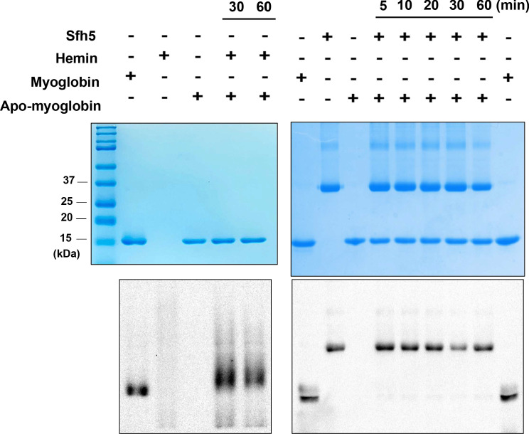
Figure 8.

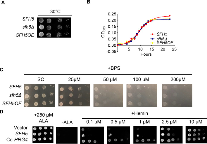
Figure 8—figure supplement 1. Sfh5 deficient cells are not compromised for major heme-requiring functions.

Figure 8—figure supplement 1.

(A) Cultures of of isogenic wild-type, sfh5Δ and Sfh5 over-expressing (OE; Sfh5 expression driven by the PMA1 promoter in the context of a genomic replacement cassette) were spotted on yeast peptone (YP) agar plates supplemented with glycerol as carbon source and incubated for 60 hr at 30°C. (B) Growth-curves for the strains described in (A) in YP-glycerol liquid media. (C) Wild-type Sfh5, sfh5Δ and SFH5OE strains grown in YPD media then harvested and washed in minimal medium plus glucose. All strains were sub-cultured in the same minimal medium for 12 hr, followed by spotting of 10-fold serial dilutions of innoculum on synthetic defined media agar plates supplemented with glucose and varied concentrations of bathophenanthroline disulfonic acid (BPS) as indicated. Plates without BPS served as control. All plates were incubated at 30°C for 36 hr and imaged. (D) A hem1Δ strain was transformed with YEp(URA3) plasmid driving galactose-inducible expression of either SFH5 or C. elegans HRG4 genes. The backbone vector plasmid served as control. After depletion of residual heme, ten-fold dilutions of cells were spotted onto minimal media plates supplemented with either α-aminolevulenic acid (ALA) or hemin as indicated. The plates were imaged after incubation at 37°C for 72 hr.
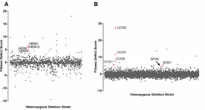
Figure 8—figure supplement 2. Specific sensitivity of Sfh5 deficient cells to oltipraz.

Figure 8—figure supplement 2.

(A) Chemogenomic profiles of the diploid yeast heterozygous deletion collection challenged with oltipraz as expressed as a log2-scaled fitness defect (FD; Lee et al., 2014). Fitness or growth defects associated with haplo-insufficiency profiling (HIP) are represented on the y-axis, and the x-axis represents statistically significant hits (p<0.05). Note the enrichment of hits directly associated with heme biosynthesis (red dots). These data are adapted from the following searchable database (http://chemogenomics.pharmacy.ubc.ca/HIPHOP/). (B) Chemogenomic profiles of the diploid yeast homozygous deletion collection challenged with oltipraz (Lee et al., 2014). The sfh5Δ homozygote is identified. Significant hits related to deficiencies in redox enzymes and Golgi trafficking components of the COG complex are also highlighted in red.