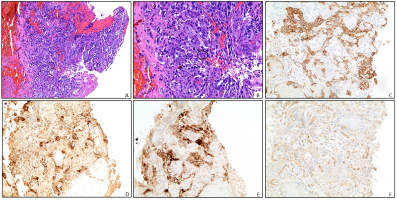
Figure 2. Pathology.
Representative pathology slides from right lung mass biopsy (A) and magnified view (B) demonstrate blood clot and necrotic tissue with rare foci of malignant cells (circled). These cells have pleomorphic nuclei, a high mitotic rate, and have an organoid to papillary architecture. There are rare cytoplasmic eosinophilic globules present. No lymphocytes are associated with the tumor. Immunohistochemical stains are as follows: (C) CK AE1/AE3 positive in tumor cells; (D) Alpha-fetoprotein (AFP) demonstrates patchy positivity; (E) GLYPICAN-3 positive; and (F) CD 117 patchy positivity. These findings, including the clinical history of an elevated AFP are consistent with a non-seminomatous germ cell tumor, yolk sac type. While no other germ cell elements are identified, the majority of this tumor is necrotic and other elements composing a mixed germ cell tumor cannot be excluded.

