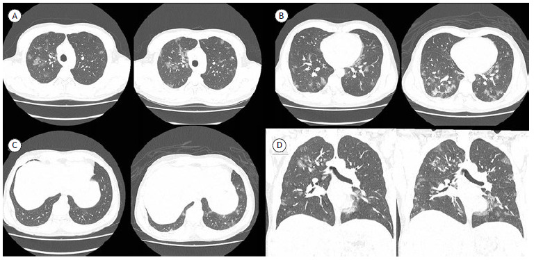
Figure 1. Axial images (in A to C) and coronal reconstructions (in D) of chest CT scans of a 44-year-old man with clinical findings suggestive of COVID-19 (fever, sore throat, and frequent dry cough), demonstrating the most commonly described pattern: numerous bilateral multifocal ground-glass opacities, associated with fine reticulation and interlobular septal thickening (crazy-paving pattern), involving various lung lobes and being predominantly peripheral in distribution in the parenchyma and a little more extensive in the posterior regions of the lower lobes. The patient had a positive RT-PCR result for COVID-19 on the day he underwent the first CT scan (images on the left in each pair) and was hospitalized. A second CT scan, which was performed three days later (images on the right in each pair) because he continued to have fever spikes and dry cough, demonstrated an increase in the number and extent of pulmonary opacities.

