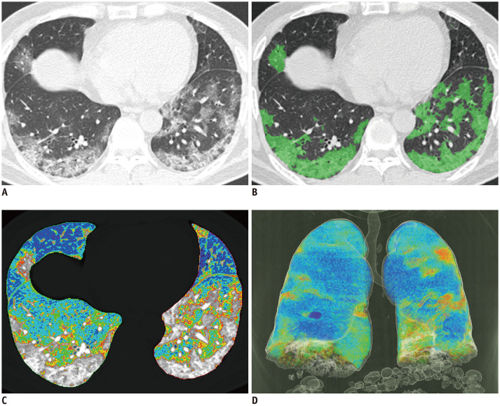
Fig. 2. COVID-19 infected 63-year-old male patient with CURB-65 score of 3.
(A) Non-enhanced axial chest CT, (B) CALP with green color-coded axial image, (C) color-coded axial image, (D) color-coded 3D reconstructed image. Color-coded regions of (C) and (D) were identified by HU according to following: purple, −1000 to −951 HU; blue, −950 to −901 HU; sky blue, −900 to −851 HU; green, −850 to −801 HU; yellow, −800 to −751 HU; and red, −750 to −701 HU. Total lung volume was 3351.0 mL, emphysema % 2.3%, NALP 65.9%, emphysema volume 77.1 mL, NAPLV 2208.3 mL, and CALPV 656.8 mL. Patient was admitted to ICU and was placed on mechanical ventilator care; however, patient died.

