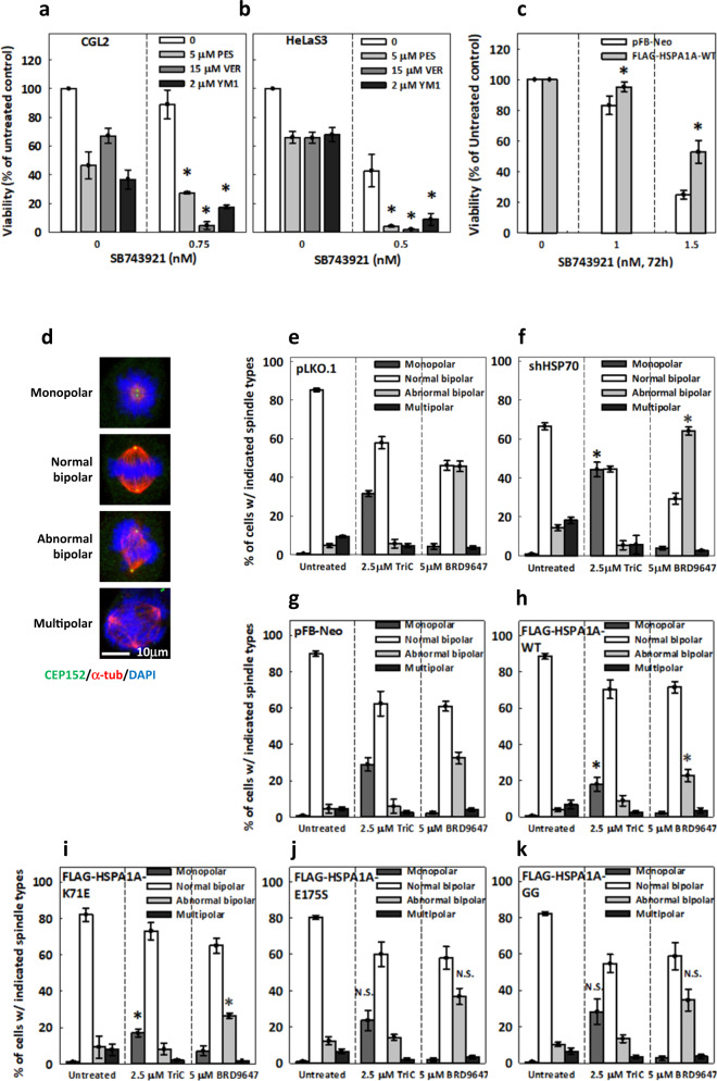
Fig. 1. HSP70 chaperone activity attenuates cell sensitivity to Eg5 inhibitors.
a–b HSP70 inhibitors enhanced the cytotoxicity of SB743921. CGL2 (a) and HeLaS3 (b) cells were incubated in medium containing SB743921 alone or in combination with HSP70 inhibitors, PES, VER or YM1, at the indicated concentrations for 72 h, after which cell viability was measured. Cell viability under HSP70 inhibitor alone was normalized to untreated cells. The mean ± SD from at least two independent experiments is shown. *p < 0.05 comparing HSP70 inhibitor-treated cells to cells without HSP70 inhibitor treatment by Student’s t test. c CGL2 cells harboring pFB-Neo empty vector or stably expressing FLAG-tagged HSP70-WT (FLAG-HSPA1A-WT) were incubated with the indicated concentration of SB743921 for 72 h before measurement of viability. Mean ± SD from at least three independent experiments is shown. *p < 0.05 comparing FLAG-HSPA1A-WT to pFB-Neo by two-way ANOVA. d Representative images of mitotic cells with the indicated types of spindle stained with CEP152 (green), α-tubulin (α-tub, red), and DAPI (blue). Cells were treated with 2.5 μM TriC or 5 μM BRD for 1 h and then fixed and immunostained to reveal mitotic spindles. e–k Percentages of cells with the indicated types of spindle are shown for the knock-down control (e, pLKO.1), HSP70-depleted (f, shHSP70), pFB-Neo-harboring (g), and FLAG-HSP70-WT (h, FLAG-HSPA1A-WT) and mutant-expressing (i–k) mitotic cells treated as indicated. At least 600 mitotic cells were counted for each experiment. The mean ± SD from at least three independent experiments is shown. *p < 0.05 comparing the respective spindle types and treatments between pLKO.1 and shHSP70 or between pFB-Neo and FLAG-HSPA1As by Student’s t test; N.S. no significance.

