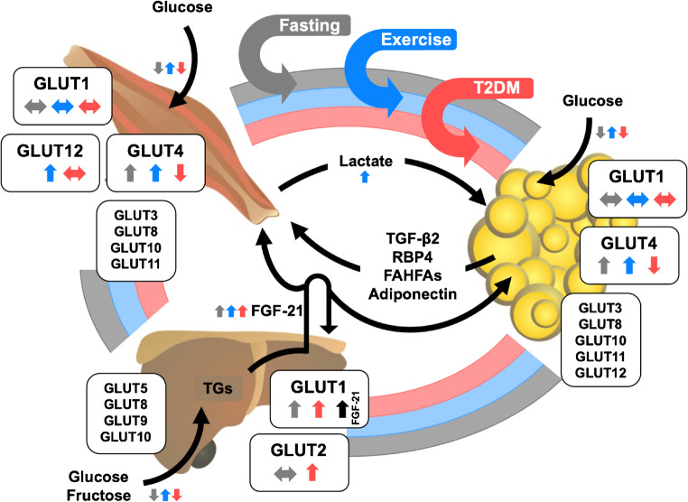
Fig. 1.
Integrative physiology of glucose transporters (GLUTs) in the liver, skeletal muscle, and adipose tissue. Expression levels of main GLUT isoforms are regulated by a diversity of metabolic stimuli including fasting and physical activity (exercise) and by certain pathophysiological conditions such as type 2 diabetes (T2DM). A complex inter-organ network is necessary to maintain whole-body energy metabolism in balance. This interaction is regulated by secretion of various factors into the circulation to facilitate tissue crosstalk. The distinct trigger mechanisms for the secretion of these factors are indicated by the respective arrow color (gray, fasting conditions; blue, exercise/physical activity; red, T2DM). In addition, the impact of these three (patho)physiological conditions on gene and/or protein expression of the diverse GLUTs as well as transport of GLUT substrates (e.g., glucose, fructose) is presented by small colored arrows next to the respective GLUT. TGs, triglycerides; FGF-21, fibroblast growth factor 21; TGF-β2, transforming growth factor β2; RBP4, retinol binding protein 4; FAHFAs, fatty acid esters of hydroxy fatty acids

