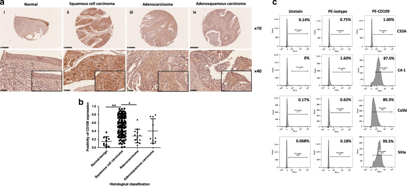
Fig. 1. Upregulation of CD109 was frequently detected in cervical squamous cell carcinoma.
a Representative immunohistochemical CD109 staining of cervical cancer tissue microarray sections: weak CD109 staining in normal cervical tissue (i), strong CD109 staining in squamous cell carcinoma (ii), mild or moderate CD109 staining in adenocarcinoma (iii) and adenosquamous cell carcinoma (iv). Magnification: upper, ×10; lower, ×40. The insets highlight regions with ×80 magnification. b Dot plot showing the distribution of CD109 immunoreactivity (presented as positivity, mean ± SD) in different histological classification groups. **P1 < 0.0001, *P2 = 0.0007, Mann–Whitney test. c CD109( + ) subpopulation in cervical cancer cell lines C33A, C4-1, CaSki and SiHa was examined by flow cytometry.

