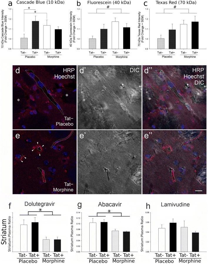
Fig. 3.
Effects of HIV-1 Tat and morphine on BBB leakiness and on antiretroviral brain concentrations. After 14 days of Tat induction, there was a significant increase in the 10 kDa (Cascade Blue®) tracer leakage into the brain in Tat + placebo as compared to Tat − placebo mice (*p < 0.05) and in Tat − mouse brains upon exposure to morphine as compared to Tat − placebo mice (*p < 0.05) (a). There was a significant main effect of morphine, resulting in reduced integrity of the BBB and increased leakage of the higher molecular weight (40 kDa and 70 kDa) tracers in morphine-exposed groups as compared to the those groups (Tat + and Tat − together) not exposed to morphine (placebo) (#p < 0.05; significant main effect of morphine) (b, c). Data represent the fold change in mean fluorescence intensity ± SEM; n = 8 Tat−/placebo, n = 6 Tat+/placebo, n = 9 Tat−/morphine, and n = 7 Tat+/morphine mice. Additionally, morphine exposure increased horseradish peroxidase (HRP) extravasation from the vasculature into the perivascular space and/or parenchyma in the striatum (d, e). HRP antigenicity was detected by indirect immunofluorescence (red) in tissue sections counterstained with Hoechst 33342 (blue) to reveal cell nuclei and visualized by differential interference contrast (DIC)-enhanced confocal microscopy. HRP extravasation into the striatal perivascular space/parenchyma was especially prevalent in morphine-exposed mice (arrowheads; left-hand panels in e versus d). The dotted lines (············) indicate the approximate edge of the capillaries/post-capillary venules; while intermittent dotted lines (· · · · · · ·) indicate the approximate edge of a partly sectioned blood vessel that appears partially outside the plane of section. The asterisks (*) indicate white matter tracts within the striatum. Representative samples from ≥ n = 4 mice per group. All images are the same magnification. Scale bar = 10 μm. Antiretroviral tissue-to-plasma ratios in striatum (f–g). Irrespective of Tat exposure, morphine significantly reduced the levels of dolutegravir (f) and abacavir (g), but not lamivudine (h), within the striatum, as compared to placebo. (* p < 0.05; main effect for morphine). Data represent the tissue-to-plasma ratios ± SEM sampled from n = 9 Tat−/placebo, n = 9 Tat+/placebo, n = 6 Tat−/morphine, and n = 8 Tat+/morphine mice. (a–h) Modified and reprinted with permission from Leibrand et al. (2019)

