Fig. 2.

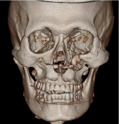
The preoperative three-dimensional computed tomography (CT) imaging. The preoperative CT imaging shows left zygomaticomaxillary complex fracture, left orbital floor fracture, right maxilla fracture, and nasal bone fracture.

The preoperative three-dimensional computed tomography (CT) imaging. The preoperative CT imaging shows left zygomaticomaxillary complex fracture, left orbital floor fracture, right maxilla fracture, and nasal bone fracture.