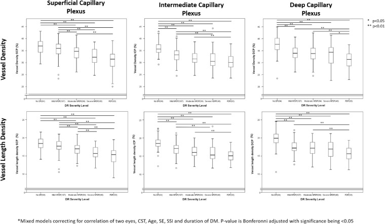
Figure 2.
Box-plots illustrating changes in all three vascular plexus with increasing diabetic retinopathy (DR) severity. Figure illustrates that with increasing DR severity, all three vascular plexuses show a decreased vessel density (VD) and vessel length density. Looking at stepwise changes, the VD and vessel length density (VLD) in intermediate and deep capillary plexuses (ICP and DCP, respectively) but not the superficial capillary plexus (SCP) is significantly lower in eyes with mild nonproliferative DR (NPDR) compared to those with no DR. With more advanced DR the DCP was not significantly lower between eyes within one DR severity while the VD/VLD SCP was significantly lower in eyes with moderate NPDR compared to those with mild NPDR and the VLD SCP was significantly lower in eyes with severe NPDR compared to those with moderate NPDR. Pair wise comparisons were performed using mixed models correcting for correlation of two eyes, CST, Age, SSI, SE and duration of DM. The P value is Bonfreroni-adjusted with significance being < 0.05.

