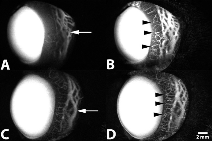
Figure 7.

Representative SA following IV administration of ICG of the left eye from a 1-year-old intact female beagle dog (A, B) and from a 2-year-old male beagle dog (C, D). (A, C) Filling within deep circumferential ciliary arteries, the intrascleral venous plexus, and the venous circle of Hovius within the lateral scleral quadrant is visualized 30 seconds after injection (arrow). (B, D) Filling of terminal ciliary arterioles, capillary beds, and venular counterparts is visualized 1 minute after injection (arrowhead).
