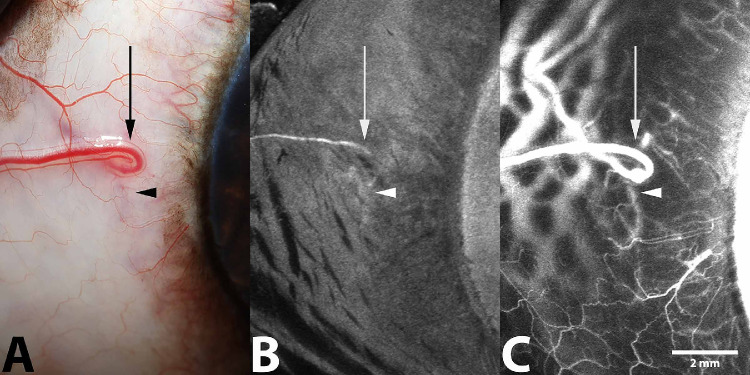
Figure 9.
Representative (A) standard color, (B) IC AA (17 minutes after ICG injection), and (C) IV SA (5 minutes after ICG injection) from the right eye of a 4-year-old male mixed-breed dog. This eye demonstrated minimal visualization of the CAHO pathways following IC administration of ICG. Outflow occurred via a component of the intrascleral plexus (arrowhead) and an episcleral vein (arrow). These components are similarly visible using standard color and IV ICG angiography.

