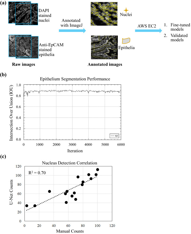
Figure 2.
U-Net based image analysis workflow. (a) Raw images of colon or small intestine with DAPI stained nuclei and anti-EpCAM stained epithelial were annotated separately by biologists using ImageJ. Annotated images were used to fine-tune or to validate U-Net models residing in AWS EC2. (b) The performance of epithelium segmentation was evaluated with Intersection Over Union (IOU). (c) The performance of nucleus detection was assessed by correlating U-Net counting with manual counting across 16 small tissue regions.

