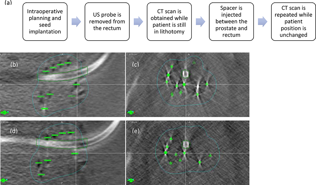
Figure 2.
(a) Steps to determine impact of spacer placement on intraprostatic seed movement Displacement of seeds within the prostate after rectal spacer placement. (b) sagittal and (c) axial CT scan images prior to rectal spacer implantation; (d) sagittal and (e) axial CT scan images after placement of rectal spacer. Note the anterior movement of seeds immediately posterior to the urethra (seed indicated by the cross‐hair) and lateral displacement of the seeds lateral to the urethra; green contour indicates 100% isodose line.

