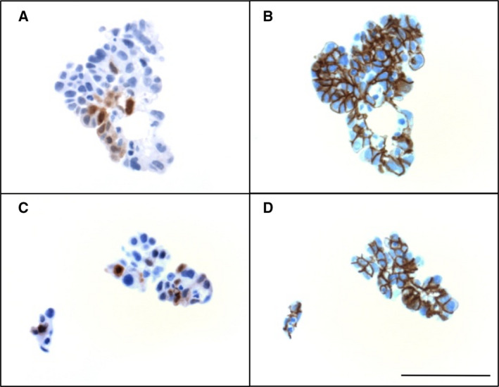
Fig. 5.

Immunohistochemical staining of rare tumor cell spheroids with calretinin‐expressing cells in ascites from 2 different HGSC patients. (A, C) Spheroids showing focal expression of calretinin. (B, D) EPCAM staining of the same spheroids showing that calretinin‐expressing cells are carcinoma cells. Scale bar: 50 µm.
