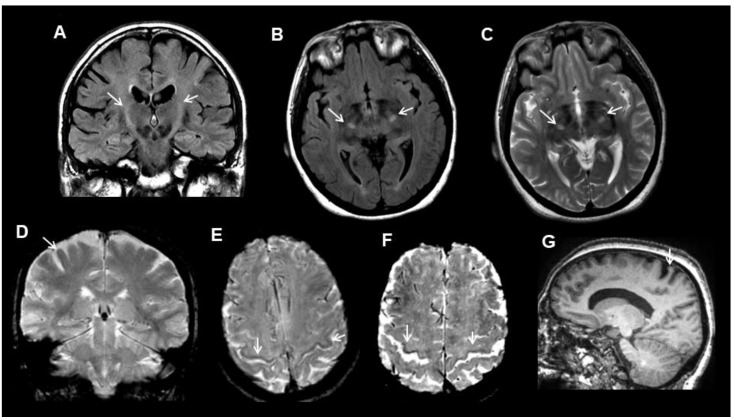
Figure 1.
Examples of the MRI (Magnetic Resonance Imaging) features (arrows) in amyotrophic lateral sclerosis (ALS) patients. (A) corticospinal tract FLAIR hyperintensity in a coronal image. (B) corticospinal tract fluid attenuated inversion recovery (FLAIR) hyperintensity in an axial image. (C) corticospinal tract T2 hyperintensity in an axial image. (D) motor cortex T2* hypointensity in a coronal image. (E) motor cortex SWI hypointensity in an axial image. (F) motor cortex SWI hypointensity and widening of the central sulcus in an axial image. (G) selective motor cortex atrophy in a sagittal-T1 image.

