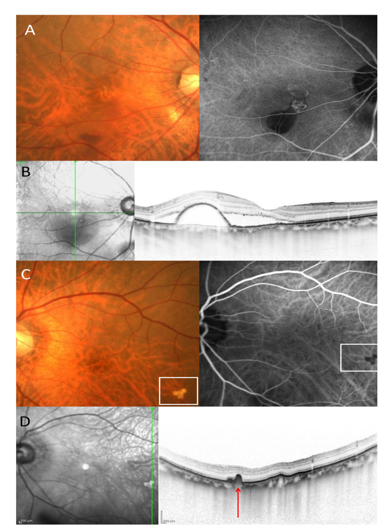
Figure 1.
An eighty-year-old male patients with polypoidal choroidal vasculopathy. (A). Color fundus photography showed hemorrhagic pigment epithelial detachment in the right eye. On the indocyanine green angiography (ICGA), polypoidal lesion and branching vascular network was seen in the right eye. (B). A vertical OCT scan through the fovea showed subretinal fluid and pigment epithelial detachment. (C). Color fundus photography showed soft drusen surrounded by a white square in the left eye. On the ICGA, the soft drusen exhibited hypofluorescent in the left eye. (D). A vertical OCT scan through the soft drusen showed retinal pigment epithelial bump (red arrow).

