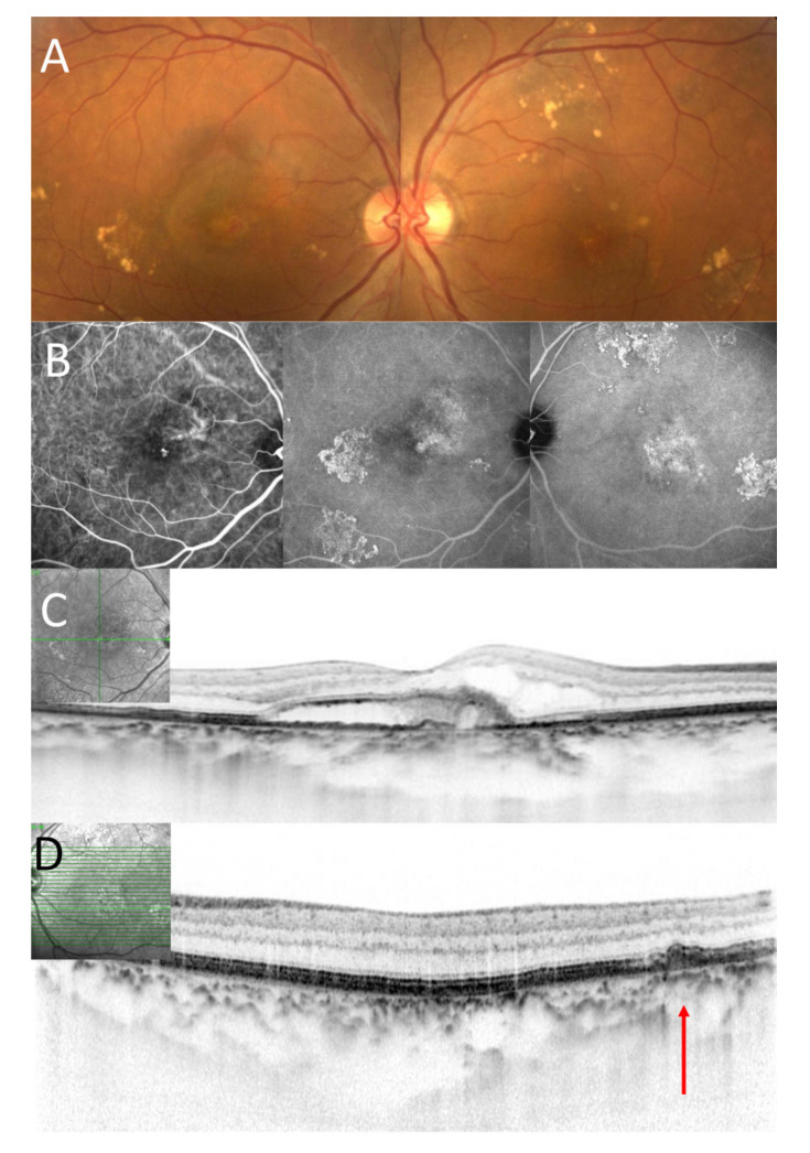
Figure 2.
A seventy-three-year-old male patient with polypoidal choroidal vasculopathy. (A). Color fundus photography showed scattered yellowish drusenoid deposits around the macular in both eyes and exudation in the macular in the right eye. (B). On the early phase of ICGA, polypoidal lesion was seen in the macula in the right eye. (Left image). On the late phase of ICGA, hyperfluorescence was seen in the area corresponding to scattered yellowish drusenoid deposits in both eyes. (Middle and left image) (C). A horizontal OCT scan through the fovea showed exudation including subretinal fluid and fibrin in the right eye. Subfoveal choroidal thickness was 375 µm in the right eye. (D). A horizontal OCT scan through the yellowish deposits showed drusenoid deposits (as indicated by a red arrow) in the left eye.

