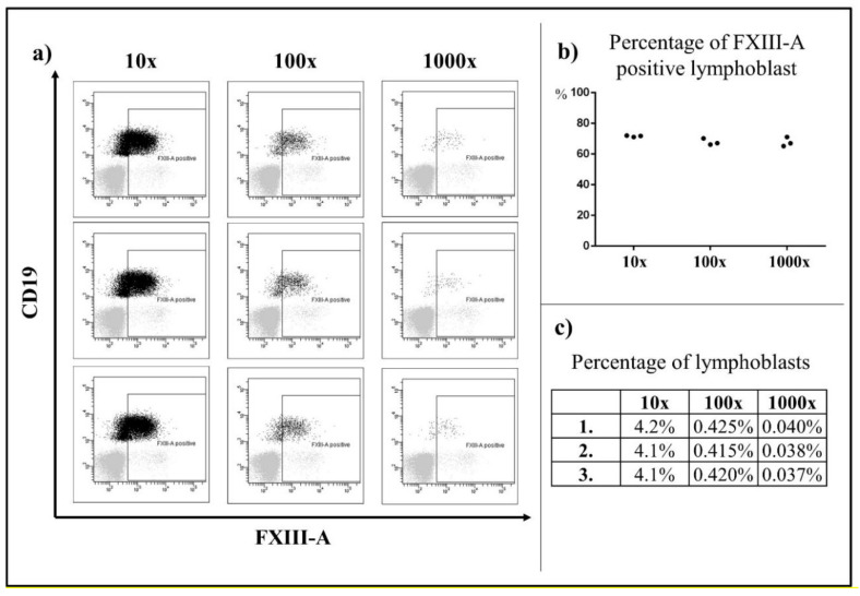
Figure 4.
Detection of FXIII-A expression on analysis of serial dilutions. Based on a three parallel serial dilutions (10×; 100×; 1000×), the percentage of FXIII-A expression in residual lymphoblasts could be clearly assessed when the percentage of lymphoblasts was above 0.04%. Black color indicates the residual lymphoblasts, grey color represents the mature lymphocytes, which were the negative control. (a) The percentages of FXIII-A positive lymphoblasts within all lymphoblasts were similar in the three parallel staining instances (b) The percentages of lymphoblasts served as the internal control of the serial dilutions (c).

