Figure 5.
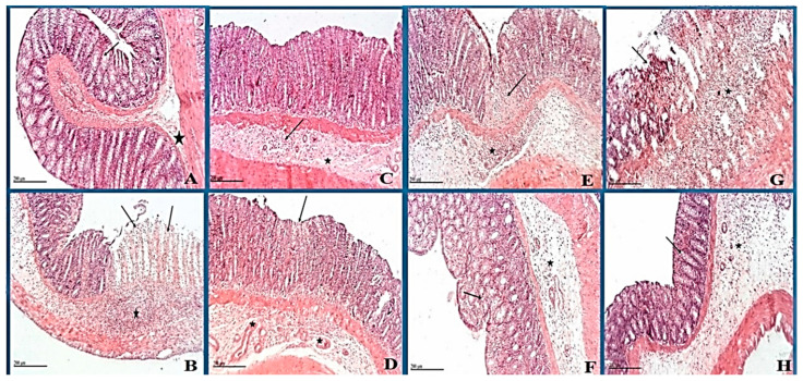
Photomicrograph of rat colon shows (A) negative control: normal histological structures of the colon with intact mucosa (star) including intestinal crypt with abundant goblet cells (arrow). (B) Positive control: extensive focal erosions and ulceration (arrow) of intestinal mucosa with necrotic tissue debris. Extensive inflammatory cells infiltration including mononuclear and polymorphonuclear cells in mucosa and submucosal layers (star) accompanied with moderate submucosal edema. (C) Dexamethasone standard: Apparent intact glandular mucosa and lining epithelium with moderate inflammatory cells infiltration in deeper mucosa and submucosal layer (arrow) accompanied with mild submucosal edema (star). (D) Nano-Ag: few focal areas of necrosis in the superficial zone of intestinal mucosa (arrow) with many intact glandular elements. Mild submucosal edema and inflammatory cells records with many dilated and/or congested submucosal blood vessels (star). (E) ASBE: tiny focal erosions and ulceration (arrow) of intestinal mucosa with necrotic tissue debris. Focal inflammatory cells infiltration including mononuclear and polymorphonuclear cells in mucosa and submucosal layers (star) accompanied with mild submucosal edema. (F) ASBE plus nano-Ag: apparent intact mucosa including glandular structures with many goblet cells. Moderate inflammatory cells infiltration in interglandular tissue (arrow) as well as submucosal layer (star) with diffuse submucosal edema. (G) ASB nano-extract: erosions and ulceration (arrow) of intestinal mucosa with necrotic tissue debris. Inflammatory cells infiltration including mononuclear and polymorphonuclear cells in mucosa and submucosal layers (star) accompanied with mild submucosal edema. (H) ASB nano extract plus nano-Ag: apparent intact mucosa including glandular structures with many goblet cells(arrow) with fewer inflammatory cells infiltration records. Focal areas of submucosal edema (star) (Normal submucosa). (H&E stain, 100 X); Scale bars for all figures (A–H) are 20 µm.

