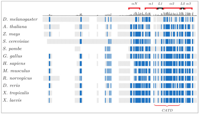
Figure 1.
CenH3 protein alignments, conservation and diversity across species. The structural elements of CenH3 proteins are illustrated, with conserved residues in blue. The histogram above the sequences shows the conserved regions: the carboxyl terminal domain and its components (L1 and α-helix) are highly preserved across eukaryotes. The shared CENP-A Targeting Domain (CATD) drives the association between proteins and centromeres [50]. Despite the variability of the amino terminal tail, this domain contains a phosphorylatable serine for CenH3 mitotic function [51]. This image is courtesy of Damien Goutte-Gattat [52].

