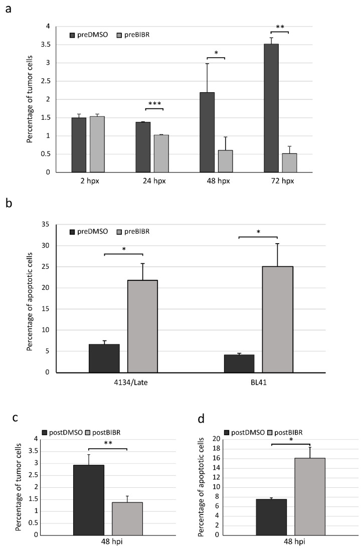
Figure 4.
Effects of BIBR on proliferation and viability of tumor cells xenografted in zebrafish embryos. (a and b) Xenografted fluorescent tumor cells, pretreated with BIBR (preBIBR) or DMSO (preDMSO), were detected by flow cytometry in enzymatically dissociated embryos. (a) Percentage of 4134/Late cells in zebrafish embryos according to time post-xenograft (hpx). Values are means and SD (bar) of three separate experiments of 10 embryos per group. (b) Cell suspensions from enzymatically dissociated embryos were processed by TUNEL assay for the detection of apoptotic cells. Histograms represent the percentage of tumor apoptotic cells (4134/Late and BL41) at 72 hpx. Values are means and SD (bar) of two separate experiments of 10 embryos per group. (c and d) Twenty-four hours after transplantation, untreated 4134/Late cells were injected with BIBR (postBIBR) or DMSO (postDMSO). Forty-eight hours after drug injection (hpi), fluorescent tumor cells were analyzed by flow cytometry in enzymatically dissociated embryos for proliferation (c) and apoptosis (d). * p < 0.05, ** p < 0.01 and *** p < 0.001.

