Figure 5.
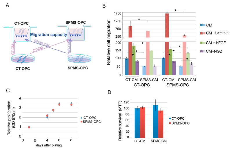
Progressive SPMS-OPC secretome shows deficient in vitro OPC migration stimulation. (A) Schematic protocol of migration assay. SPMS- and CT-derived early OPC-like cells were culture in fasting media, and conditional media (CM) were recovered after 24 h and added to the lower well of transwell chambers. SPMS- and CT-derived early OPC-like cells were plated on upper wells of transwell chambers, and 24 h later, migrated cells on the lower membrane surface were fixed, crystal violet stained, and quantified by optical density (OD 540) values of stained cell extracts. (B) Quantification of cell migration (OD 540) under CT- or SPMS-derived CM with indicated stimulating factors (laminin, bFGF, or NG2) relative to the CT-OPC-like sample under CT CM stimulation. Error bars represent means ± SEM (n = 6 independent experiments with early OPC-like cell lines from four different SPMS and three CT donors) * p ≤ 0.05 by unpaired Student’s t-test. (C) SPMS- and CT-derived early OPC-like cells present similar proliferation rates. A total of 25,000 OPC-like cells were cultured with NIM/bFGF/purmorphamine for proliferation/viability MTT (3-(4,5-dimethylthiazol-2-yl)-2,5-diphenyltetrazolium bromide) assay. Cells were recovered 1, 4, 5, 6, and 8 days after and measured at 570 nm absorbance, reflecting the number of viable cells (MTT assay). Average relative absorbance ± SEM (relative to day 1) is represented (n = 3 independent experiments with early OPC-like cell lines from four different SPMS and three CT donors). (D) CT and SPMS-derived OPC-like cells were cultured for 24 h in CM as in Figure 5A, and after 48 h, the number of viable cells (MTT assay) was measured. Average relative absorbance ± SEM is represented (n = 3 independent experiments with OPC-like cell lines from four different SPMS and three CT donors).

