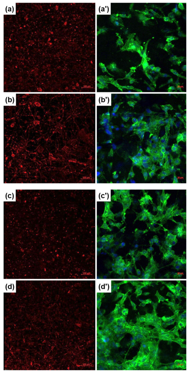
Figure 7.

Fluorescence microscopy images recorded on fetal osteoblasts cultured for 24/48 h of: (a,a’) PCL-Mg-1, (b,b’) PCL-Mg-5, (c,c’) PCL-Sr-1 and (d,d’) PCL-Sr-5 scaffolds, achieved with: (a–d) CellTracker Red CMTPX or (a’–d’) Fluorescein Phalloidin with DAPI.
