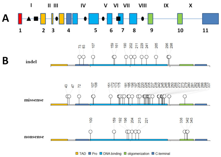
Figure 1.
Schematic representation of the genomic locus of the TP53 gene and its protein sequence with marked gene alterations present in osteosarcoma patients derived samples. (Panel A) represents mutations (●), deletions (■) and translocations (∆) locations in particular introns. Roman numerals refer to introns, Arabic numerals refer to exons. (Panel B) represents p53 protein primary structure with marked domains and amino acids number of mutations (indels, missense, nonsense). TAD—transactivation domain; Pro—proline-rich region; DNA binding—core domain which can bind DNA; oligomerization—oligomerization domain. Based on data from [17,18,29,62,63].

