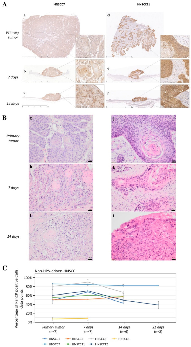
Figure 2.
Morphology and PanCK-immunohistochemistry of two non-HPV-driven 3D-OTCs. An anti-PanCK antibody was used to visualize the amount and spatial distribution of PanCK-positive cancer cells (brown signal). For visualization of cellular integrity, details in 16-fold higher magnification are shown for each sample (A). Scale Bar in images with lower magnification: 4 mm, and higher magnisfication: 100 µm. (B) H/E staining of the according samples. Scale bar: 20 µm. (C) Mean values of tumor cell proportion of all primaries and 3D-OTCs on day 7, 14, and 21 of all non-HPV-driven HNSCC. Error bars indicate standard errors of the mean. Abbreviations: PanCK, pan-cytokeratin; HPV, human papillomavirus; 3D-OTC, 3D organotypic co-culture; H/E, hematoxylin and eosin. HNSCC, head and neck squamous cell carcinoma.

