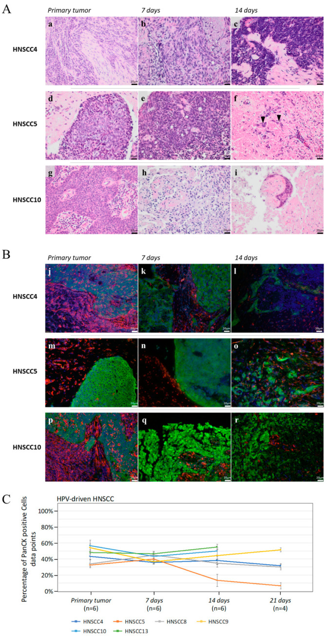
Figure 7.
(A) H/E staining of three different HPV-driven 3D-HNSCC-OTC (b–f,h,i) and corresponding primaries (a,d,g). HNSCC4 maintains its tumorous morphology. HNSCC5 shows few remaining tumor cells on day 14 (f: black arrowheads) and HNSCC10 presents an altered morphology. Scale Bar: 20 µm. (B) Co-immunofluorescence staining with an anti-PanCK-antibody (green), an anti-vimentin-antibody (red), and DAPI (blue) of the samples presented in (A), confirming the observations. Scale Bar: 20 µm. (C) Mean values of tumor cell proportion of all primaries and 3D-OTCs on day 7, 14, and 21 of all HPV-driven HNSCC. Error bars indicate standard errors of the mean. Abbreviations: H/E, hematoxylin/eosin; HPV, human papillomavirus; 3D-OTC, 3D organotypic co-culture; PanCK, pan-cytokeratin.

