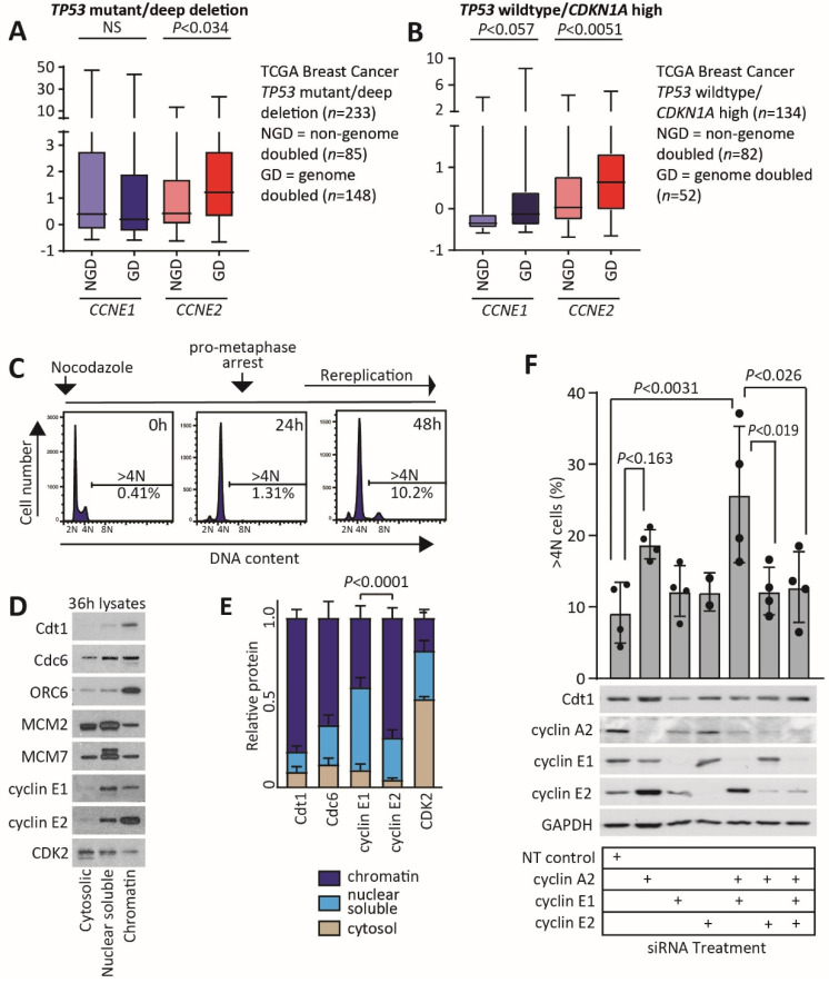
Figure 6.
Cyclin E2 is associated with genome doubling in p53 wildtype breast cancers. (A) Relative CCNE1 and CCNE2 expression was determined across the TCGA breast cancer dataset and compared between non-genome doubled (NGD) and genome doubled (GD) cancers with TP53 mutation/deep deletion. Data were analysed by Welch’s t-tests. (B) Relative CCNE1 and CCNE2 expression was determined across the TCGA breast cancer dataset and compared between non-genome doubled (NGD) and genome doubled (GD) cancers with wildtype TP53 status, high TP53 expression and high CDKN1A expression. Data were analysed by Welch’s t-tests. (C) Schematic of nocodazole-mediated arrest in MCF-7 cells and effect on cell cycle distribution at 0 h, 24 h and 48 h, determined by propidium iodide staining of cells and detection of >4N cells by flow cytometry. N = sets of chromosomes. (D) Cytosolic, nuclear soluble and chromatin lysates were collected at 36 h following nocodazole addition, and western blotted for the preRC proteins Cdt1, Cdc6, ORC6, MCM2, MCM7 and for cyclin E1, cyclin E2 and CDK2. (E) The relative proportion of protein in each cell fraction was quantitated by ImageJ, and compared with a chi-squared test. (F) MCF-7 cells transfected with siRNAs to cyclin A2, cyclin E1, cyclin E2 and non-targeting control were blocked at pro-metaphase with 50 ng/mL nocodazole, and cells were collected 48 h later for analysis for >4N cells by flow cytometry and by western blotting. Treatments were compared by one-way ANOVA, with Tukey’s multiple comparisons test. The uncropped western blot figure in Figure S11.

