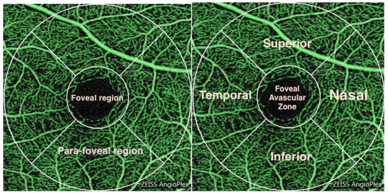
Figure 1.
Pictorial representation of the macular vasculature. The vessels in a 3 mm radius annulus centred at the fovea were measured. Division of the annulus into foveal and para-foveal regions is shown along with further division into superior, inferior, nasal and temporal quadrants. The radius of the aggregated foveal/parafoveal region is 3 mm whilst the foveal region has a 1mm radius.

