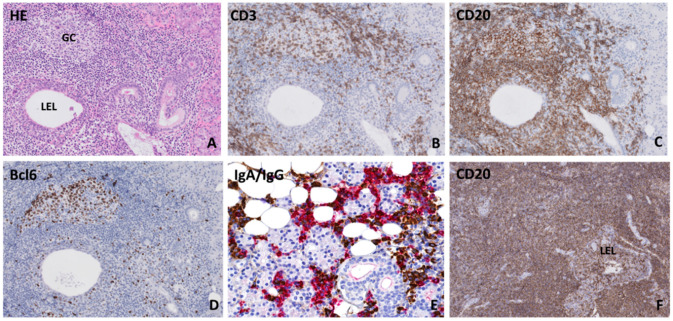
Figure 1.
Histopathological features in parotid salivary glands of primary Sjögren’s syndrome patients (A) Lymphocytic infiltrate located around a hyperplastic striated duct (lymphoepithelial lesion: LEL) without obstructed lumen. Both (B) CD3+ T-lymphocytes and (C) CD20+ B-lymphocytes are present in the periductal infiltrate and within the ductal epithelium. (D) Presence of a germinal center, which was revealed by the presence of a cluster of ≥5 adjacent Bcl6-positive cells within a focus [28]. (E) Immunoglobulin A (IgA) (red) and immunoglobulin G (IgG) (brown) staining shows a plasma cell shift towards IgG plasma cells. (F) Salivary gland mucosa associated lymphoid tissue (MALT) lymphoma biopsy, which shows a diffuse CD20+ B-lymphocytic infiltrate around lymphoepithelial lesions in the absence of normal salivary gland parenchyma.

