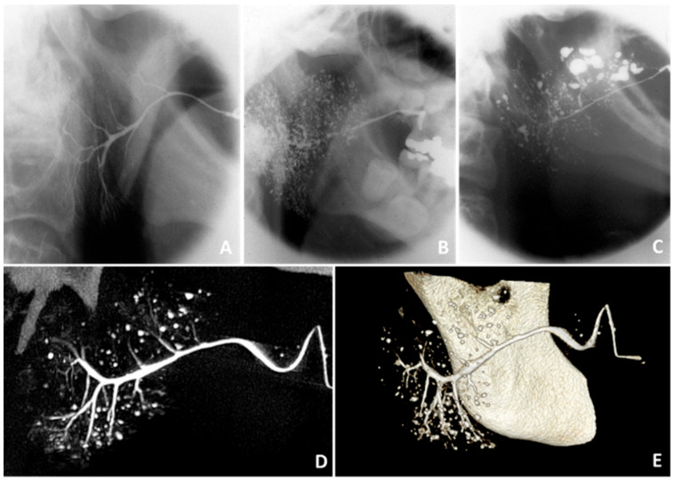
Figure 2.
Findings on sialography. Sialographies of the parotid gland showing (A) no abnormalities in a healthy subject, (B) punctate/globular sialectasis in a pSS patient, and (C) globular/cavitary sialectasis in a pSS patient [29]. (D) Two-dimensional sialo-CBCT image and (E) three-dimensional sialo-CBCT image of the parotid gland of a pSS patient, showing normal width of the primary duct, moderate scarcity of ductal branches, and numerous diverse sialectasis. Thanks to Prof. D.J. Aframian and Dr. C. Nadler and colleagues who provided the sialo-CBCT images.

