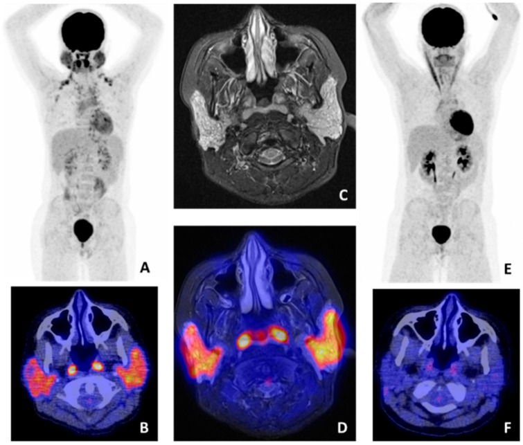
Figure 3.
18F-fluorodeoxyglucose (FDG) positron emmison tomography/computed tomography (PET/CT) and magnetic resonance imaging (MRI) findings in a pSS patient with salivary gland mucosa associated lymphoid tissue (MALT) lymphoma. (A) Whole-body FDG-PET showing high heterogeneous FDG uptake in both parotid and submandibular glands. No other pathological lesions were found (axillary and clavicular regions with increased uptake represent brown fat). (B) FDG-PET/CT image showing pathological uptake in the parotid glands and physiological uptake in the tonsils. (C) MRI stir sequence showing a pathological, heterogeneous aspect of both parotid glands. (D) Manually fused FDG-PET/MRI image, showing pathological uptake in the parotid glands and physiological uptake in the tonsils. (E) Whole-body FDG-PET and (F) FDG-PET/CT image after treatment, showing no pathological uptake in the parotid glands, indicating complete remission.

