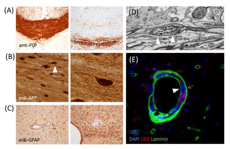
Figure 2.
(A) Shows anti-proteolipid protein staining of the control corpus callosum (left) and corpus callosum of a cuprizone-intoxicated (right) mouse. (B) Shows a section stained with the anti-amyloid precursor protein of a mouse intoxicated with cuprizone. The axonal spheroid highlighted by the arrowhead is displayed on the right site at a higher magnification. (C) Shows the anti-glial fibrillary acidic protein-stained sections of a cuprizone-experimental autoimmune encephalomyelitis (EAE) mouse [41]. The image on the left shows moderate, and the image on the right shows severe, astrogliosis. (D) Shows the ultrastructure of an axonal spheroid. (E) Shows a perivascular inflammatory infiltrate stained with anti-CD3 and anti-laminin to label T-lymphocytes and basement membranes, respectively. The arrow highlights the astrocyte basement membrane, while the arrowhead highlights the endothelial basement membrane.

