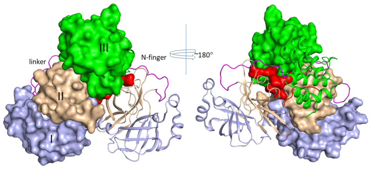
Figure 2.
Structure of SARS-CoV-2 3CLpro. The N-terminal seven residues (N-finger), domains I, II, III, and the linker of domains II and III of both protomers are shown in red, light blue, wheat, green, and purple, respectively. The linker in the two protomers is shown in ribbon mode. Other domains in one protomer are shown in surface mode except the linker region, and corresponding domains in the other protomer are shown in ribbon mode. The structure (PDB ID 6Y2G) is used in this figure.

